")
 (1)")
")
 (1)")
 Description
 Specifications
 Applications
 Pictures
 Downloads
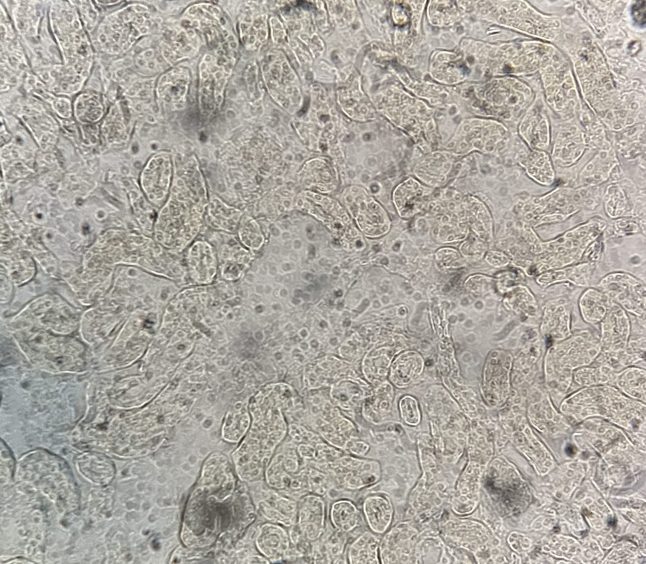
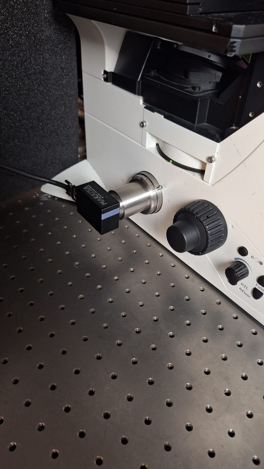
MAKATEA camera, quantitative phase imaging made easy
MAKATEA is an affordable camera based on QLSI (Quadriwave Lateral Shearing Interferometry), a technique that enables the mapping of wavefront profiles with exceptional sensitivity (<1 nm) and high spatial resolution (approaching the diffraction limit). MAKATEA is extremely compact (60x40x26mm) and was specifically built for seamless integration into quantitative phase imaging systems. Using a simple C-Mount adapter, MAKATEA can be effortlessly installed on any optical microscope camera port. Using the most simple transmitted light mode with white light, MAKATEA outputs both intensity and phase images live with a refresh rate of 10- 15 fps without using any specific phase contrast rings or objectives.
Quantitative phase imaging (QPI) is a microscopy technique that measures the phase shift of light as it passes through a sample. It is a label-free method that can be used to image :
- Living cells and tissues: neurons and neurites, organelles, vesicles, bacteria, eukaryotic cells, algae, yeast, archaea, viruses, etc.
- Nanophotonic structures: nanoparticles, 2D materials, metasurfaces, nanowires, and more.
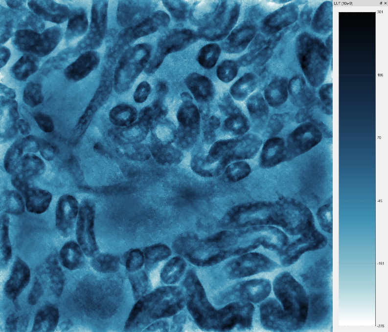
Transmitted light image with regular camera QPI image acquired with MAKATEA
The images above were acquired at the Axiom Optics lab using a Leica DMI 8000 widefield microscope in transmitted light mode (white light) and a 20x objective. In this example, the transmitted light image shows very little contrast as cells are transparent. However because of local changes of index of refraction and thickness variations, these cells have a very significant impact on the phase of the light. The QPI image provided by MAKATEA maps those changes and shows excellent contrast. Because it is quantitative, it can be further processed to extract physical properties of cells (ex: dry mass).
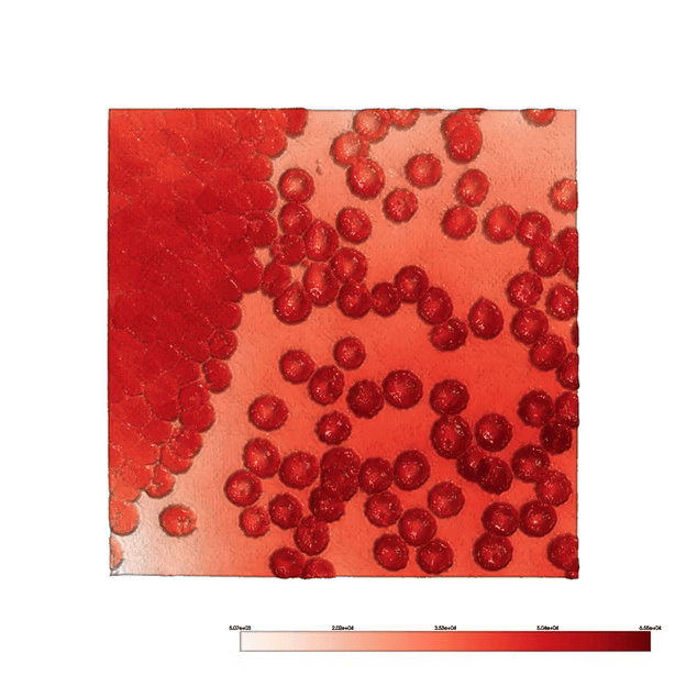
The MAKATEA camera is particularly effective for single-cell dry mass measurement and analysis from just a single image acquisition. Over its lifetime, a cell’s dry mass—comprising the total mass of cellular components excluding water—undergoes significant changes influenced by metabolic and structural activities. By analyzing these variations, valuable insights can be gained into cellular dynamics such as the cell cycle, responses to stimuli or drugs, and metabolic activity. This information is instrumental for studying both individual cells and cell populations.
Quantitative Phase Imaging Applications:
- Determination of biomass in individual live cells with precision below a picogram and measurement of dry mass density (pg/µm²).
- Imaging of birefringence properties.
- Visualization of the complex optical polarizability of nanoparticles.
- Characterization of the complex refractive index in graphene and other two-dimensional materials.
- Temperature mapping at the microscopic scale.
- Profiling of wavefronts generated by metasurfaces.
- Single-exposure phase-fluorescence imaging.
- Assessment of surface roughness and topography with an accuracy of λ/1000.
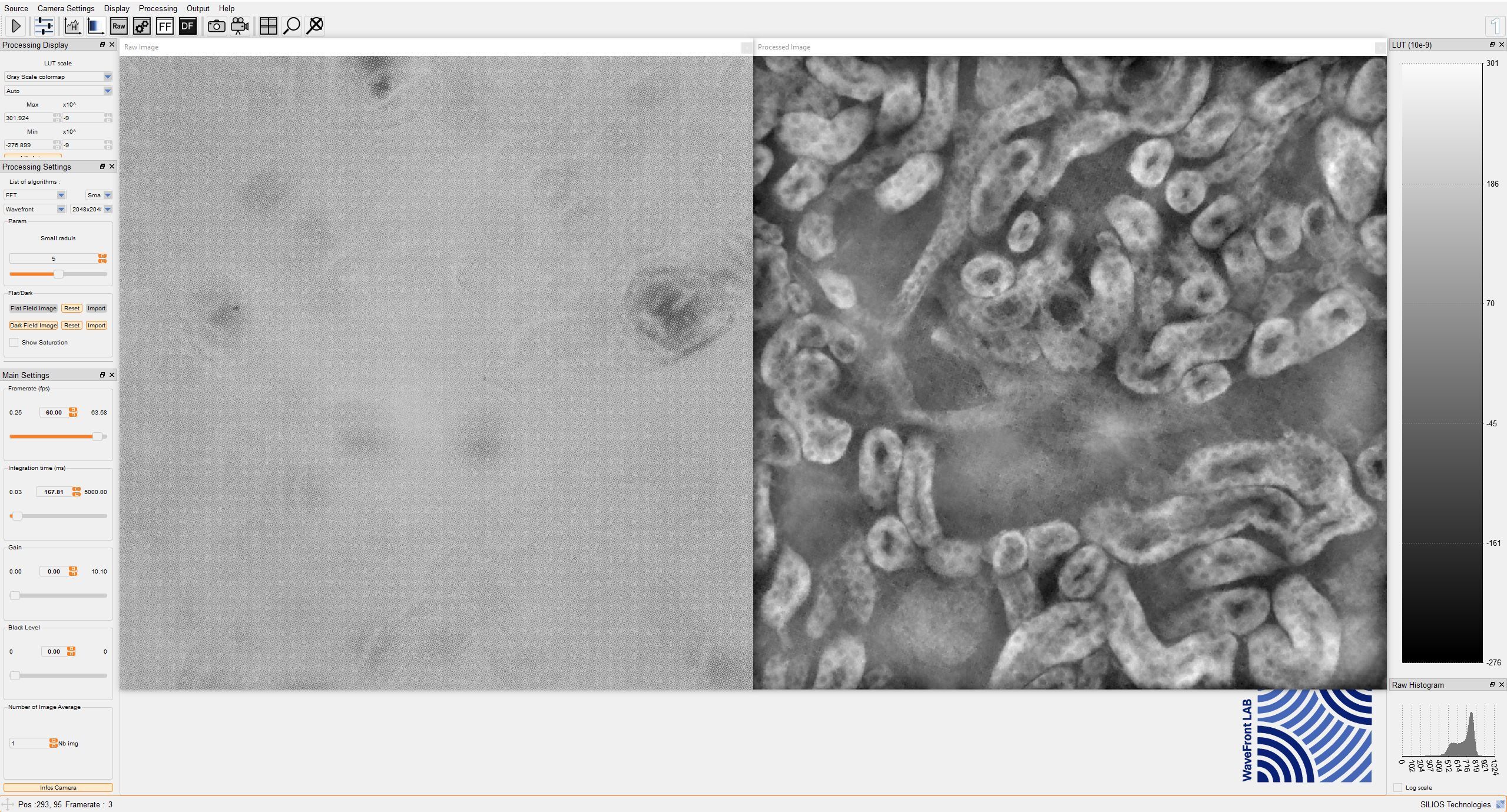
Software Development Kit (SDK)
WaveFront LAB is the SDK designed for SILIOS WFI cameras for quantitative phase imaging. It offers the following capabilities:
- Visualization of raw, wavefront, gradient, and transmittance images.
- Display of both regular and restored full-size images.
- Two calculation modes: a fast method based on Fourier Transformation and a more precise approach utilizing the Sylvester equation.
WaveFront LAB operates in real-time acquisition mode but also supports single-image or multi-image processing in “replay mode.” It is compatible with all SILIOS cameras and automatically identifies connected devices using their serial numbers. The specific spectral data corresponding to the WFI sensor are applied during processing. WaveFront LAB is provided free of charge with the purchase of any multispectral WFI camera. The SDK supports development in C/C++ and Python.
View the manufacturer’s page here: https://www.silios.com/makatea-cam
View more Quantitative Phase Imaging Cameras, click here.